
maple
Canadian Medical Alliance for the Preservation of the Lower Extremity
Charcot Neuroarthropathy
Charcot neuroathropathy is a condition where, in cases of
significant abnormal nerve function known as neuropathy,
the bones and joints of the foot break down and develop
complex fractures, fragmentation and dislocations that
results in a collapse of the arch and normal anatomical
structure of the foot.
These complex fractures result in deformity, altered function,
and as a result, a greater risk of ulcers, infections, and
amputation.
As Charcot neuroarthropathy is caused by a sensory loss of neuropathy, we should mention that the topic of neuropathy is discussed at greater length here. But to summarize, some patients, particularly diabetics, may develop abnormal nerve function that can result in a sensory loss--an inability to feel pain. It is those with sensory loss at risk to develop Charcot neuroarthropathy.
History of Charcot Neuroarthropathy
Charcot (pronounced "Shark-oh", with the accent on the 2nd syllable)
neuroarthropathy was named after Jean Martin Charcot (1825-1893),
a French physician considered today as the father of neurology.
It was he who first identified the disease, and because Charcot
recognized that this condition is associated with nerves ("neuro"-)
and joint ("arthro-") pathology ("-pathy"), it bears the name
Charcot neuroarthropathy.
Charcot neuroarthropathy is also known as Charcot arthropathy, Charcot
joint disease, Charcot disease, or simply, Charcot.
Charcot was so important in the field of neurology that at least 15 medical
conditions, signs or features have been named after him. Besides Charcot
neuroarthropathy, Charcot-Marie-Tooth disease was named for Charcot,
and Lou Gehrig's disease was originally named after Charcot, as well.
Besides being named for these conditions, Charcot produced landmark
papers on multiple personality disorders, syphilis, stroke, epilepsy,
multiple sclerosis and Parkinson's disease. In fact, it was Charcot, himself,
bestowed the name Parkinson's disease to the English physician James
Parkinson.
Equally impressive are the names of those who trained under Charcot, like the neurologist Joseph Babinski (after whom the famous foot reflex is named), neurologist - psychologist Pierre Janet (who, among other things, first described the behavior known as 'la belle indifférence', discussed on the page on arterial ulcers), Alfred Binet (who developed the first practical IQ test) and Georges Gilles de la Tourette (after whom Charcot named Tourette's syndrome).
Charcot's most famous student, however, was the psychiatrist, Sigmund Freud. Freud translated Charcot's lectures into German and used the lectures on hysteria to form the foundations of psychoanalysis. Freud also named his son, Jean-Martin, after Charcot.
Charcot identified this condition of complex fractures,
bone destruction and joint degeneration in patients with
advanced syphilis in 1868. At the time, there were no
antibiotics, and syphilis would commonly progress to the
point of nerve loss.
Leprosy patients, too, frequently developed the condition
in Charcot's day, but today is rarely seen because of the
development of antibiotics.
Today, however, with antibiotics able to treat syphilis and
leprosy patients, very few patients with those conditions
progress to this stage.
However, diabetic patients, who often didn't live long
enough to develop Charcot neuroarthropathy in Charcot's
time, are living so long that this group has become, far
and away, the most common group that develop the nerve
disorder (neuropathy) that causes Charcot.
However, the condition of Charcot neuroarthropathy may
be seen in a variety of other conditions, including chronic
alcoholism, drug toxicity, trauma, syringomyelia, severe
spinal compression, multiple sclerosis, rheumatoid arthritis,
psoriatic arthritis, and a hereditary insensitivity to pain.
How common is Charcot disease?
Fortunately, Charcot is not particularly common. Approximately one Canadian in ten will develop diabetes, and just one diabetic in 700 will develop Charcot (1).
However, this means family practitioners come across a Charcot patient relatively rarely, and often may not recognize it when it appears or know how to treat it. This can lead to greater deformity and complications.
"To learn how to treat a disease,
one must learn how to recognize it."
--Jean-Martin Charcot
As a clinical example, the patient pictured here
presented to the hospital with a red, hot, swollen
foot. There was no open wound. But because of
the redness, heat and edema, it was assumed he
had an infection.
The patient was placed on IV antibiotics for 8 weeks,
followed by several more weeks of oral antibiotics.
However, the redness and heat and swelling did not
resolve. The radiographs below show why. Note
the significant fracturing, dislocation, arch collapse,
and deformity caused by weight bearing on the foot.
This is all consistent with an acute Charcot.
How does Charcot develop?
Charcot is found in those who have a significant loss of sensation to the foot and,
typically, good circulation.
The loss of sensation, combined with an accumulation of small, repetitive micro-
traumas to the foot leads to Charcot. The body cannot feel these microtraumas,
and the foot does not have time to heal from them. Fractures and degenerative
changes result.
With this said, larger traumas are also associated with the development of Charcot.
Ankle fractures with a surgical repair is a common example of a precipitating factor,
as seen in these examples. So one must be watchful for Charcot changes in a
neuropathic foot, but particularly so in one that has recently undergone major foot
trauma or foot surgery.
Trauma creates inflammation. And inflammation stimulates
the cells that break down bone, cells known as (osteoclasts).
And because patients cannot feel the damage occurring
in their feet, they continue to walk, create more damage,
which triggers more inflammation, which stimulates more
osteoclasts. Thus, the downward spiral.
In diabetic patients, there may be another factor at play.
The increased sugar content (glycosylation) of the strong,
white connective tissues (like ligaments, capsule, fascia,
periosteum, and tendon) that hold the foot together weaken
these tissues. Over time, the body weight stretches these
connective tissues, creating misalignments in bone structure.
Diagnosis
Because of all the bone changes shown above, it may be assumed that x-rays are a primary tool to assess the foot for Charcot. And they are. The radiographs above are classic for Charcot. However, s-rays may not show much early. Sometimes you can't even see a fracture. There may be just a wispy, cotton-candy-like bone callus at the edge of a bone--part of the body's attempt to heal (below right).
Sometimes there's not even this to go on.
Because Charcot patients cannot feel pain, their first symptoms are usually an
unexplained swelling in the foot. They may notice redness and an increased
temperature to the foot (below).
Because there is no pain, patients often ignore these symptoms for weeks or months, and the foot begins to degenerate, and becomes more and more misshapen--usually wider with a lower arch.
It is important not to ignore one's early symptoms, even if you can't feel it. As Charcot once said,
"Symptoms, are in reality nothing
but a cry from suffering organs."
-- Jean-Martin Charcot
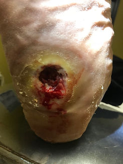
As the foot becomes more deformed, patients may develop neuropathic ulcerations (holes in the skin). However, in Charcot, the ulcers often develop in unusual locations that normally bear no weight, such as the arch of the foot. Unlike most diabetic and neuropathic ulcerations in the foot, it is classically the midfoot that is affected by Charcot. The rearfoot and ankle are next, followed by the forefoot (metatarsals and toes).
The classic signs of Charcot--swelling, warmth and redness--can, of course, be found in other medical conditions, such as trauma, infection and gout. And because many physicians don't see Charcot regularly, the condition is often misdiagnosed and mistreated, leading to significant deformity that can make ambulation difficult and lead to more ulcerations.
In the case below, Charcot was missed early and under-treated Note the significant ankle fracture and misalignment of the leg to the foot. The body weight drives the leg bone (tibia) down into the ground, the foot is pushed to the side, and a significant ulcer develops and degenerative arthritis develops through the midfoot. Deformities like these usually require complete reconstruction. This despite the fact that the reconstruction, itself, may sometimes precipitate more Charcot.
So Charcot is often diagnosed by physical examination. And x-rays are nearly always used to assess the condition and monitor changes over time. You'll often see fractures of bone and the obliteration of joints and normal architecture.
In this first group of films, note
the multiple, complex fractures--
like a small explosion took place
in the foot.
As with these examples, the
midfoot is the classic location for
Charcot neuroarthropathy to
develop.
It's often difficult to see normal
joint spaces, or tell which
fragment belongs to what bone.
With this fracturing and fragmentation come deformities.
And the deformities that develop through Charcot are
permanent.
So the sooner the condition is properly identified, the sooner
treatment can be begun, and the sooner any deformity can be
arrested.
To the right, note the misalignment of the bones.
The red lines represent bone bisections and should be relatively
straight to each other.
Note widening of the foot as the body weight collapses the
arch and the bones splay.
Fragments of bone sometimes look like they're being spat out, or discarded by the foot (below).
The degree of pathology and deformity can be substantial (below). The foot may become non-functional. Surgical reconstruction or amputation may be required.
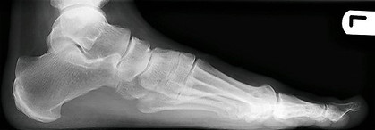
One of the most common misalignment issues seen in Charcot is a collapsed arch. Here you see an x-ray of a normal foot (below left) compared to a foot where the arch has collapsed (below right).
When the arch collapses enough from the body weight
that the bones of the midfoot reach the ground (as seen
to the right), the mid-arch ulcerations unique to Charcot
become likely.
When the arch collapses so much it becomes inverted--a
"rocker-bottom" foot, with no flat surface to walk on.
Even without the risks of ulcers and infections from the
bony prominence, walking on the large bony mass on the
bottom of the foot can be quite difficult.
Besides x-ray, other imaging (CT, MRI) may be useful early in the
disease, to see the extent of damage present. These technologies
give nice, cross-sectional views into the foot, as seen to the right.
As you see here, there are multiple fragments of bone breaking
down, making it difficult to differentiate which piece belongs to
what bone.
Regular bone scans show bone activity, but they are not specific
enough to differentiate between different possible diagnoses such
as infection which can cause that increased bone activity.
However, white blood cell bone scans may be useful to rule out a
bone infection, which is the most common differential diagnosis.
Foot specialists and those who see a lot of diabetic wounds can
usually diagnose the condition much earlier than physicians who
do not specialize in the foot.
Differential Diagnoses (What else could it be?)
Approximately 20% of diabetics will eventually have an ulcer. And many of these will become infected at some point, a condition that brings with it, swelling, redness and an increase in temperature. These are the same symptoms most Charcot patients experience. And because Charcot is much less common that a diabetic foot infection, there is a natural tendency by physicians, particularly physicians who do not see Charcot frequently, to assume any red, hot swollen foot is infected.
Radiographs of bone infection (osteomyelitis) can mimic Charcot, too, as both conditions may result in bone and joint destruction.
The problem is that antibiotics will not treat Charcot. And when Charcot is missed, significant bone and joint destruction may develop, leading in many cases to frank deformity.
Another common condition that brings with it a red, hot, swollen foot
is gout. This, too, is a common mis-diagnosis, and it, too, can lead to
significant deformity while the wrong condition is being treated.
Blood clots (DVT) can also create a hot, swollen leg that can mimic
Charcot.
Trauma, such as a midfoot or ankle fracture, can also cause a swollen,
deformed foot that can look like Charcot.
Posterior Tibial Tendon Dysfunction (PTTD), seen to the right, is a
condition where the major muscle and tendon that holds up the arch
and pushes the body forward in gait no longer functions. It commonly
leads to a collapsed arch and swollen foot and ankle, mimicking
Charcot.
It's been estimated that 25% of Charcot patients have been initially
misdiagnosed or undiagnosed (2), but in the experience of the author
of this web page, this number could be much higher.
How do you treat Charcot?
The single most important thing one needs to do with Charcot is
getting all pressure off the wound. If you're allowed to walk, you're
allowing the deformity to progress.
Luckily, removing pressure from ulcers to get them to heal is also the
way to treat Charcot. In fact, we have to be even more aggressive with
removing pressure to allow the bones the opportunity to consolidate
and heal.
We do this by total non-weight bearing. This may mean a wheel chair,
crutches or a knee-walker (right). And ideally, this should be coupled
with a total contact cast (TCC).
The TCC allows for stability, particularly if the patient is non-compliant
and does bear weight. And it's quite effective in reducing edema.
The TCC is usually changed in 3-4 days after the first application to
adjust for changes in swelling within the leg, this is changed weekly.
We monitor the improvement of the condition by watching for signs of consolidation and bone healing on regular x-rays. We monitor the temperature of the foot. A warm foot is still a sign of danger. We want the temperature of the Charcot foot to return to the temperature of the unaffected foot. And when ulcers are present along with Charcot, we manage them to closure.
It should be noted that the TCC, and immobilization
in general, does carry some risks. The patient may
have a heightened risk of blood clots. There is a
greater risk of a fall. There can be loss of muscle
tone and bone density (cast sickness). But this
must be balanced against the devastating
consequences of an untreated Charcot.
X-rays are typically taken regularly to watch
for further destruction of bone and evidence
of healing.
Monitoring the temperature of the foot is also
helpful. When the foot is still hot, it is likely still
active Charcot, still in the process of destruction.
As the foot heals, weight bearing may be considered.
One may be able to bear weight sooner with the use of a
total contact cast (below left), a custom-made CROW boot
(CROW=Charcot Restraint Orthotic Walker, below middle, which
is removable and more convenient, or an over-the-counter diabetic
offloading boot (below right), which is significantly less expensive, but
less effective at immobilization.
Considering the degree of deformity and dysfunction severe Charcot can cause, however, it is better to be overly cautious (and have the patient stay off the foot longer) than underly cautious (and allow early walking that can lead to more bone and joint destruction and deformity.
This is particularly true if the treating physician is less experienced, the foot is particularly unstable, or if there is a question as to the degree of resolution present.
Long-Term Management of the Charcot Foot
Once the Charcot is resolved and the foot is healed, the deformity that Charcot creates needs to be accommodated. The choice depends upon how early the Charcot was diagnosed and how quickly and how aggressive treatment was initiated.
If little to no deformity exists, standard footwear, such as a high-quality running shoe, may be possible.
If the foot is modestly deformed (a wider, larger foot), a wider running shoe (such as New Balance, which comes in different widths).
With moderate deformity on the bottom of the foot
(plantarly), a custom orthotic (near right) may be
required to offload small areas of prominent bone.
On the far right we see a patient's own, home-made
attempt to get weight off an abnormal bony prominence.
However, having a professionally-made device, preferably
by a podiatrist who specializes in this sort of deformity,
will almost always produce better results.
Below is a $500 insert that was made by a store-front, retail operation for a patient with an active Charcot and a non-resolving ulcer of over 1-year duration. It offers no support (below left and more importantly, no offloading whatsoever. This highlights the need to have a specialist involved in your care.
Another option are over-the-counter, offloading shoes (below left) and over-the-counter, offloading sandals (below center), both of which come with some system of thick, modifiable innersoles (below right). In some cases, this can be used long term to offload an ulcer and / or to house a wider, thicker, larger Charcot foot. This is especially useful in those who cannot afford more expensive custom-made options.
In cases of greater deformity, a custom shoe may be required. Below center, a patient preparing casted to offload a prominent region of bone on the outside of the sole of the foot.
A custom boot (below right) may be required if additional ankle stability is required.
One should wait until the swelling subsides before making a custom shoe.
In more severe cases, surgery may also be necessary to resolve bony abnormalities that predispose the patient to future ulcerations and infections or to provide stability for bony structures left in unstable orientations.
Keep in mind, however, surgery can set of more Charcot.
And keep in mind, once you've had Charcot, additional episodes of Charcot are more likely, even without surgery. Even on the opposing limb, Charcot is more likely to develop once you've had Charcot. One must be ever vigilant for future bouts of Charcot.
The earlier you diagnose Charcot, the better, not only in terms of resolving the issue, but to prevent the complications that occur with deformity and predisposition to ulcers, infection and dysfunction.
1) https://www.ahni.com/Specialties/Foot+and+Ankle/Articles/Common+Disorders/Charcot+Joint+Disease+-+Neuroarthropathy.html
2) Myerson MS, Henderson MR, Saxby T, Short KW. Management of midfoot diabetic Neuropathy. Foot Ankle Int 1994; 15(5):233-241.


Jean-Martin Charcot (1825-1893)


"Given the choice between the experience
of pain and nothing, I would choose pain."
--WIlliam Faulkner


Charcot's famous hospital, Salpêtrière, in Paris. Salpêtrière was once the site of a gunpowder factory. (Hence the name Salpêtrière, the French word for "Saltpeter," one of the ingredients of gunpowder.) Louis XIV built the current building to house the poor and mentally ill, but it evolved into the world's largest hospital. F1 driver Michael Schumacher was treated here, as was French president Jacques Chirac and Prince Ranier of Monaco. Josephine Baker and Princess Diana died here. It is now the teaching hospital for the Sorbonne


Body Weight


Stages of Charcot Foot
Phase 1 The acute, active, developmental phase of Charcot, with new fragmentation, dislocation and degeneration, where the foot is red, hot and swollen.
Phase 2 The coalescence phase, where the fractures begin to come together and heal, swelling diminishes and the temperature of the foot drops
Phase 3 The consolidation phase, where the bones are healed, the foot is stable, and the temperature returns to normal. Deformity and inflexibility may persist.


To return to the top of this page,
click on the maple leaf to the right.
This page written by Dr. S A Schumacher
Podiatric Surgeon, Surrey, British Columbia Canada
All clinical photographs on this page are provided by Dr. S A Schumacher. They may be reproduced for educational purposes with attribution to:
Dr. S A Schumacher, Surrey, BC Canada,
and a link to this website, www.CanadianMAPLE.org.

